TAVI
TAVI
We have been performing TAVI procedures in Derriford Hospital since 2008 and have performed over 3000 procedures during this time with excellent results. Mr Dalrymple-Hay and Mr Lloyd are the 2 surgeons who perform these procedures and we work very closely with our Cardiology colleagues and our SCP teams throughout your workup and procedure. We have a robust Multidisciplinary Team to assess all patients referred for surgery.
What is a TAVI?
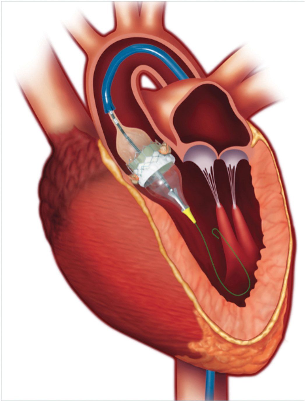
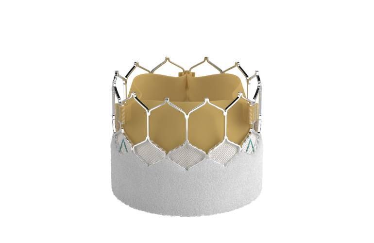
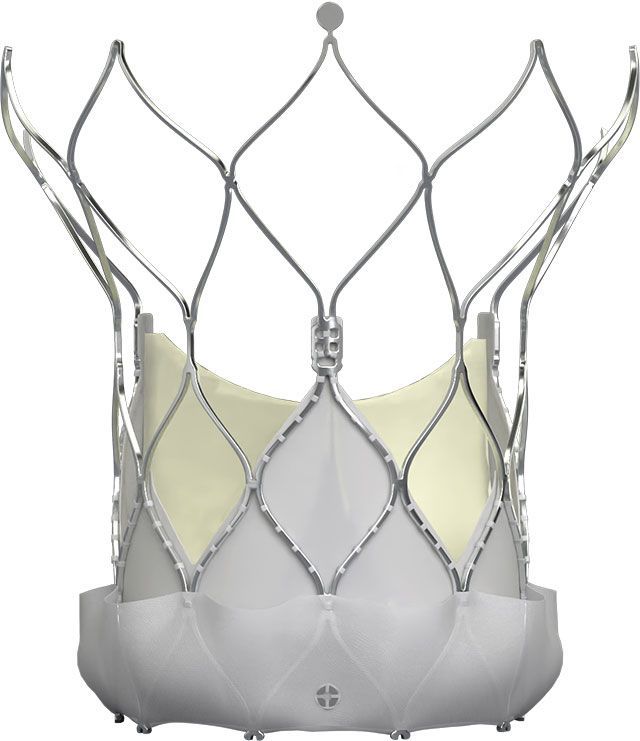
TAVI is short for trans-catheter aortic valve implantation, and is a way of improving how your heart works without us having to remove your own narrowed valve. It involves putting an artificial valve into your heart. The valve is made up of three ‘leaflets’ (made of material derived from cows) which control blood flow. The valve is inside a metal cage called a stent, which holds the new valve in position.
You do not need open heart surgery to have this procedure. Instead, the valve is inserted into the heart through a thin tube (known as a catheter) which is put into the body through a small cut in either your groin or your chest.
The procedure takes up to two hours and is carried out under local or general anaesthetic. Because this is a less invasive procedure with a shorter anaesthetic than open-heart surgery, it means that a person considered too high a risk for open heart surgery may face a lower risk with this new procedure.
About the Procedure
There are a number of ways that the valve can be implanted:
- Transfemoral: This is performed through a small cut into one of your groin arteries and is performed usually under local anaesthesia. Recovery is usually 24-48hrs.
- Trans Axillary: This is performed through a small cut near your shoulder under short general anaesthesia. Recovery is usually 24-48hrs.
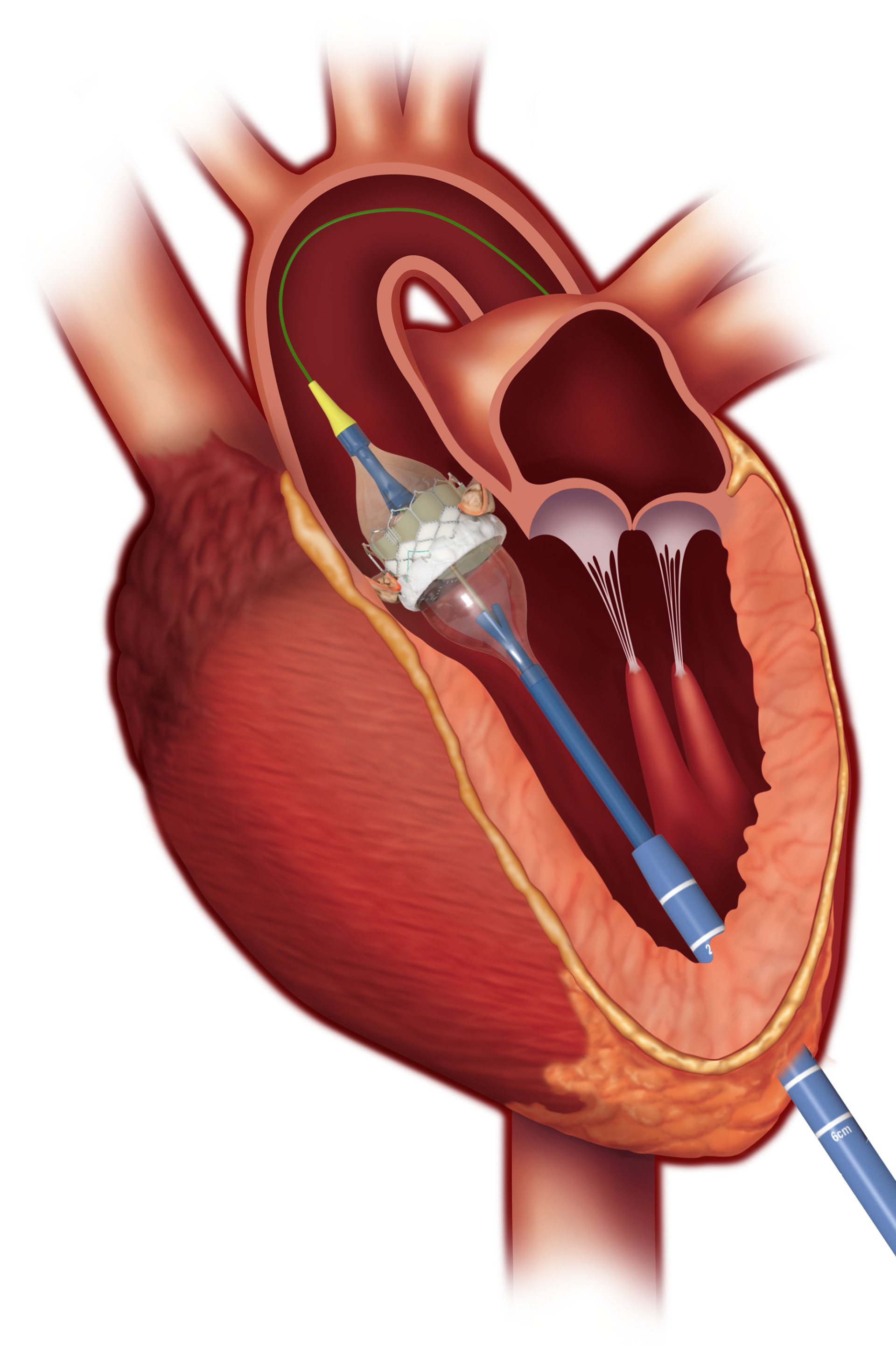
- Transapical: This is performed through a small incision in your chest wall and is performed under general anaesthesia. Recovery is usually a littler longer over 3-4 days in hospital.
We will also put a pacing wire into a vein in your leg. This wire acts as a pacemaker, allowing us to temporarily speed up your heart at key points in the procedure. This means that less blood is passing through your aortic valve, enabling us to ensure that the artificial valve is positioned correctly.
We will put your new valve in place by making a small cut in your groin and/or your chest. If we use your groin for the procedure, this is referred to as a trans-femoral procedure. If we use your arm vessel it is called transaxillary and if we use an incision directly through your chest, we call it a trans-apical procedure.
Your cardiac surgeon and cardiologist will decide which is best for you based on the results of your TAVI work up tests, and the cut will be repaired at the end of the procedure. When you wake up you may have puncture sites in your groin from the pacing wire and tubes (sheaths) used to get the images we require for positioning the valve.
Through these sheaths, we will pass a fine tube (catheter) to your heart. Before your artificial valve can be implanted, it is carefully crimped (compressed) and mounted onto the balloon. We then pass the balloon and mounted valve through the cut in your groin, arm or chest and put it in place across your aortic valve. Once it is in place, your artificial valve is expanded by the balloon so that it fits across your existing valve, holding it open permanently. This procedure will improve how well your heart works without you having to have your own diseased aortic valve removed.
What happens after the procedure?
Initially you will be transferred to the Recovery Unit in theatres so that you can be closely monitored. You will then be transferred to either the coronary care unit (CCU) or back to the ward.
During your stay in hospital, you will also have the following tests:
- A chest x-ray.
- Urine analysis.
- Routine blood tests to check how well your kidneys and liver are working, to make sure you do not have a blood infection, to check that your blood can clot properly, and to make sure you have enough haemoglobin, which carries oxygen around in your blood.
- An electrocardiogram (ECG).
- A trans-thoracic echocardiogram. You will have this just before you leave hospital after your procedure. It involves having an ultrasound probe placed on your chest so it can record images of your heart.
You are likely to be in hospital for approximately 2-3 days after your procedure.


